Anatomy and physiology laboratories provide hands-on experience‚ bridging theoretical knowledge with practical application. Labs enable exploration of biological structures and physiological processes‚ fostering curiosity and critical thinking. Safety protocols and ethical considerations are emphasized to ensure responsible practices. Students engage in investigative activities‚ using microscopes‚ dissection tools‚ and modern technology to enhance learning. This foundational experience prepares individuals for advanced studies and professional careers in healthcare and science.
1.1 Importance of Lab Work in Understanding Anatomy and Physiology
The importance of lab work in understanding anatomy and physiology lies in its ability to bridge theoretical knowledge with practical application. Hands-on experiences‚ such as microscopic examinations and dissections‚ allow students to observe and study biological structures firsthand. Lab activities enhance comprehension of complex physiological processes and foster critical thinking. By engaging in investigative procedures‚ students develop essential skills in scientific inquiry and data interpretation. These experiences are crucial for building a strong foundation in anatomy and physiology‚ preparing learners for careers in healthcare and research.
1.2 Overview of Lab Safety and Ethical Considerations
Lab safety and ethical considerations are paramount in anatomy and physiology laboratories. Proper handling of chemicals‚ biological specimens‚ and equipment is essential to prevent accidents. Students must adhere to safety protocols‚ such as wearing protective gear and following emergency procedures. Ethical practices include respecting cadaveric specimens and minimizing environmental impact through proper waste disposal. Compliance with lab regulations ensures a safe and responsible learning environment‚ fostering accountability and respect for scientific practices.
Cellular and Tissue Studies
Cellular and tissue studies form the foundation of understanding human anatomy and physiology. Microscopic examinations reveal cellular structures‚ while histological techniques prepare tissues for detailed analysis. Identifying epithelial‚ connective‚ muscle‚ and nervous tissues enhances comprehension of their roles in maintaining bodily functions.
2.1 Microscopic Examination of Cells and Tissues
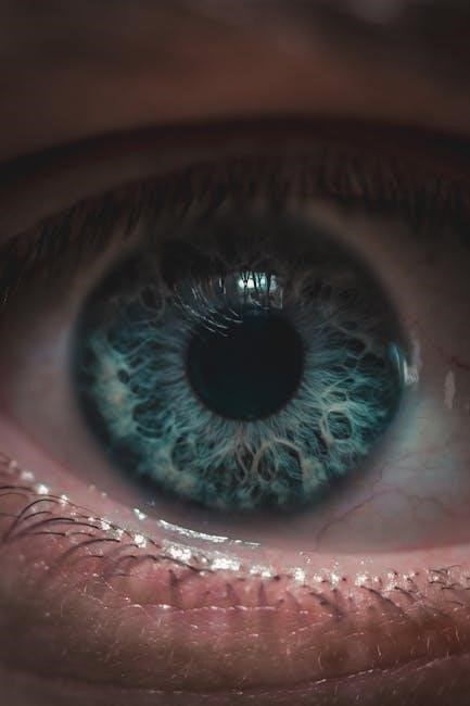
Microscopic examination is a cornerstone of cellular and tissue studies‚ enabling detailed observation of cellular structures and tissue organization. Using light microscopes‚ students analyze prepared slides to identify key features such as cell shapes‚ nuclei‚ and organelles. Staining techniques enhance visibility‚ distinguishing between different tissue types like epithelial‚ connective‚ and muscle tissues. This hands-on approach helps students correlate microscopic structures with their physiological functions‚ fostering a deeper understanding of human anatomy and pathology.
2.2 Histological Preparation Techniques
Histological preparation involves several steps to transform tissues into slides for microscopic examination. Fixation stabilizes cellular structures‚ while dehydration and embedding prepare tissues for sectioning. Staining enhances contrast‚ allowing differentiation of cellular components. Techniques like paraffin wax embedding and frozen sectioning are commonly used. Proper preparation ensures high-quality images‚ enabling accurate identification of tissue types and cellular details; These methods are essential for both educational and research purposes in anatomy and physiology.
2.3 Identifying Epithelial‚ Connective‚ Muscle‚ and Nervous Tissues
Epithelial tissues are identified by their layered‚ tightly packed cells forming barriers or lining surfaces. Connective tissues‚ rich in collagen‚ provide support and include bone‚ cartilage‚ and blood. Muscle tissues are characterized by elongated‚ contractile cells‚ while nervous tissues contain neurons and glial cells for signaling. Histological slides‚ often stained with H&E‚ reveal distinct structures. Proper identification requires understanding cellular arrangements and specialized staining techniques. These skills are foundational for analyzing tissue samples in anatomy and physiology labs.
Organ Systems Exploration
Organ systems exploration involves studying the structure and function of major systems like skeletal‚ muscular‚ nervous‚ and circulatory. Labs use dissection and microscopy to examine tissues and organs‚ providing insights into system interactions and physiological processes. This hands-on approach helps integrate theoretical knowledge with practical observations‚ enhancing understanding of human anatomy and physiology.
3.1 Skeletal System: Bone Structure and Joint Movements
The skeletal system consists of bones‚ cartilage‚ and ligaments‚ providing structural support and protection. Bones are classified into long‚ short‚ flat‚ and irregular types‚ each with unique functions. Lab activities include examining bone tissue under microscopes to identify compact and cancellous bone. Joint movements are studied to understand synovial‚ cartilaginous‚ and fibrous joints‚ emphasizing their roles in mobility. Practical exercises‚ such as dissection and X-ray analysis‚ help students visualize bone architecture and joint mechanics‚ reinforcing concepts of skeletal anatomy and physiology.
3.2 Muscular System: Types of Muscles and Their Functions
The muscular system comprises three types of muscles: skeletal‚ smooth‚ and cardiac. Skeletal muscles‚ attached to bones‚ enable voluntary movements and maintain posture. Smooth muscles‚ found in internal organs‚ facilitate involuntary actions like digestion. Cardiac muscle‚ exclusive to the heart‚ ensures continuous pumping of blood. Lab exercises involve identifying muscle tissues under microscopes and observing their contraction mechanisms. Understanding muscle functions and their roles in movement‚ support‚ and internal processes is crucial for comprehending human physiology and diagnosing related disorders.
3.3 Nervous System: Brain‚ Spinal Cord‚ and Peripheral Nerves
The nervous system is divided into the central nervous system (CNS)‚ including the brain and spinal cord‚ and the peripheral nervous system (PNS). The brain controls higher functions like thought‚ memory‚ and voluntary movements. The spinal cord facilitates reflexes and serves as a pathway for nerve signals. Peripheral nerves transmit sensory and motor signals between the CNS and the body. Lab activities include dissecting nerve tissues and studying nerve impulse conduction‚ essential for understanding nervous system functions and disorders.
3.4 Circulatory System: Heart Structure and Blood Vessel Types
The circulatory system consists of the heart and blood vessels‚ responsible for transporting blood throughout the body. The heart is a muscular organ with four chambers: two atria and two ventricles. Arteries carry oxygenated blood away from the heart‚ while veins return deoxygenated blood. Capillaries facilitate gas and nutrient exchange. In lab‚ students examine heart structures‚ measure blood pressure‚ and study blood vessel histology to understand their functions and roles in maintaining homeostasis.
3.5 Respiratory System: Lung Structure and Breathing Mechanisms
The respiratory system is essential for exchanging oxygen and carbon dioxide through breathing. The lungs are paired organs protected by the rib cage‚ with lobes divided into bronchopulmonary segments. Air enters via the trachea‚ branching into bronchi and bronchioles‚ reaching alveoli for gas exchange. Lab activities include studying lung histology‚ measuring respiratory rates‚ and understanding mechanisms like inhalation and exhalation‚ vital for maintaining cellular oxygenation and overall bodily functions.
3.6 Digestive System: Gastrointestinal Tract and Accessory Organs
The digestive system processes food into nutrients‚ beginning with the mouth and esophagus‚ followed by the stomach‚ small intestine‚ and large intestine. Accessory organs like the liver‚ pancreas‚ and gallbladder produce enzymes and bile to aid digestion. Lab exercises involve examining histological slides of intestinal tissues‚ observing enzyme activity‚ and simulating digestive processes. Understanding these mechanisms is crucial for grasping nutrient absorption and the role of each organ in maintaining overall health and metabolism.
3.7 Urinary System: Kidney Function and Urine Formation
The urinary system primarily functions through the kidneys‚ which filter blood to produce urine. Nephrons‚ the functional units‚ perform filtration‚ reabsorption‚ and secretion to regulate electrolytes and fluid balance. Lab activities include examining kidney histology‚ observing urinalysis‚ and simulating filtration processes. Understanding these mechanisms is essential for appreciating how the kidneys maintain homeostasis and overall bodily functions through precise regulation of waste removal and electrolyte balance.
3.8 Endocrine System: Glands and Hormone Secretion
The endocrine system consists of glands that produce and secrete hormones regulating various bodily functions. Key glands include the pituitary‚ thyroid‚ adrenal‚ pancreas‚ and gonads. Each gland synthesizes specific hormones‚ such as insulin‚ thyroxine‚ and adrenaline‚ which control processes like metabolism‚ growth‚ and stress response. Lab exercises involve studying gland histology‚ hormone assays‚ and simulations of feedback mechanisms. These activities enhance understanding of endocrine regulation and its role in maintaining homeostasis and overall health.
3.9 Reproductive System: Male and Female Anatomy
The reproductive system involves structures essential for sexual reproduction and fertility. In males‚ key organs include the testes‚ epididymis‚ vas deferens‚ seminal vesicles‚ prostate‚ and penis‚ functioning in sperm production and delivery. In females‚ the ovaries‚ fallopian tubes‚ uterus‚ cervix‚ and vagina support egg production‚ fertilization‚ and pregnancy. Laboratory studies include anatomical dissections‚ histological examinations‚ and functional analyses to understand reproductive processes and their significance in human biology and health.

Practical Laboratory Exercises
Practical lab exercises involve hands-on activities like dissections‚ physiological measurements‚ and equipment usage. Students gain skills in data collection‚ analysis‚ and applying theoretical knowledge to real-world scenarios safely.
4.1 Dissection Techniques and Tools
Dissection techniques require precision and care to examine anatomical structures. Essential tools include scalpels‚ forceps‚ and dissecting microscopes. Students learn proper handling and safety protocols to avoid accidents. Effective techniques involve making precise incisions and gently exposing tissues for observation. Understanding the anatomy of specimens enhances comprehension of physiological functions. Proper tool maintenance and cleanup are emphasized to ensure a safe and efficient learning environment. These skills are fundamental for future careers in healthcare and scientific research.
4.2 Measuring Physiological Parameters: Heart Rate‚ Blood Pressure‚ and Respiratory Rate
Measuring physiological parameters is crucial for understanding human function. Heart rate is typically assessed using pulse palpation or ECG‚ while blood pressure is measured with a sphygmomanometer. Respiratory rate is counted by observing chest movements or using a spirometer. These measurements provide insights into cardiovascular and respiratory health. Normal ranges vary‚ but averages are 60-100 beats per minute for heart rate‚ 90-120/60-80 mmHg for blood pressure‚ and 12-20 breaths per minute for respiratory rate. Accurate techniques ensure reliable data collection.
4.3 Using Laboratory Equipment: Microscopes‚ pH Meters‚ and Electrical Stimulation Devices
Microscopes are essential for examining cellular and tissue structures‚ providing magnified views of biological samples. pH meters measure acidity levels in solutions‚ crucial for understanding physiological environments. Electrical stimulation devices apply controlled currents to observe muscle and nerve responses‚ aiding in the study of motor function. Proper calibration and handling of these tools ensure accurate and reliable experimental results. Students learn to operate this equipment to enhance their understanding of anatomical and physiological processes in a controlled laboratory setting.
Histology and Microscopy
Histology involves the study of tissue structure under microscopes‚ utilizing stains to enhance visibility of cellular details. This technique is fundamental for understanding tissue organization and function.
5;1 Preparing and Staining Tissue Samples
Preparing tissue samples involves fixation‚ sectioning‚ and staining to maintain structure and enhance visibility. Fixation preserves tissues‚ while sectioning creates thin slices for microscopy. Staining‚ such as hematoxylin and eosin (H&E)‚ highlights cellular details. Proper techniques ensure accurate observations‚ aiding in tissue identification and study. Safety protocols are essential when handling chemicals and biological specimens to prevent hazards. This process is critical for histological analysis‚ enabling students to explore tissue composition and function effectively.
5.2 Identifying Cellular Structures Under a Microscope
Microscopic examination allows detailed observation of cellular structures‚ such as cell membranes‚ nuclei‚ and organelles. Proper staining enhances visibility‚ distinguishing components like mitochondria and ribosomes. Students learn to focus and adjust microscope settings for clear images. Identifying these structures aids in understanding cellular function and tissue composition. Regular practice improves proficiency in recognizing normal and abnormal cellular features‚ which is essential for histological analysis and diagnosing conditions. This skill is foundational for advanced studies in anatomy and physiology.
5.3 Common Histological Stains and Their Applications
Common histological stains‚ such as hematoxylin and eosin (H&E)‚ are widely used to differentiate tissue components. H&E highlights nuclei and cytoplasm‚ aiding in identifying cell types and structures. Special stains like Masson’s trichrome reveal collagen fibers‚ while Gram stain distinguishes bacterial cell walls. These techniques enhance microscopic observations‚ enabling precise tissue analysis. Proper staining is critical for diagnosing pathological conditions and understanding normal tissue architecture. Mastery of staining methods is essential for accurate histological interpretations in research and clinical settings.
Physiology of Motor Control
The basal ganglia regulate motor functions‚ requiring dopamine for proper input processing. Neural pathways coordinate movements‚ while reflex actions enable rapid responses‚ ensuring precise and adaptive motor control mechanisms.
6.1 Role of the Basal Ganglia in Motor Function
The basal ganglia‚ a group of brain nuclei‚ play a crucial role in motor function by regulating voluntary movements and coordinating muscle activity. They act as a relay station‚ processing motor signals between the cortex and thalamus. Proper functioning requires dopamine release at the input nuclei‚ ensuring smooth and controlled movements. Dysfunction in this system can lead to disorders such as Parkinson’s disease‚ characterized by tremors and rigidity. Understanding the basal ganglia’s mechanisms is essential for developing treatments for motor-related neurological conditions.
6.2 Dopamine’s Role in Movement Regulation
Dopamine plays a critical role in regulating movement by modulating neural pathways within the basal ganglia. It facilitates smooth‚ coordinated motor actions by balancing excitatory and inhibitory signals. Dopamine release in the striatum‚ the input nucleus of the basal ganglia‚ is essential for initiating voluntary movements. Imbalances in dopamine levels‚ such as deficiencies‚ lead to movement disorders like Parkinson’s disease‚ characterized by tremors‚ rigidity‚ and bradykinesia. Understanding dopamine’s mechanisms is vital for developing therapies to restore normal motor function in such conditions.
6.3 Neural Pathways and Reflex Actions
Neural pathways are sequences of neurons that transmit signals‚ enabling communication between different parts of the body. These pathways are essential for controlling involuntary and voluntary actions. Reflex actions‚ such as withdrawing a hand from heat‚ occur rapidly without conscious thought. They involve sensory neurons detecting stimuli‚ interneurons processing signals‚ and motor neurons triggering responses. Understanding these pathways and reflex mechanisms is crucial for comprehending how the nervous system coordinates and regulates bodily functions‚ ensuring survival and maintaining homeostasis.

Plant Anatomy and Root Development
Plant anatomy focuses on the study of root structures‚ revealing their architectural traits and functional roles in water and nutrient absorption. Root development is influenced by genetic factors‚ environmental conditions‚ and evolutionary adaptations. Understanding root anatomy aids in improving crop yields and stress tolerance‚ making it vital for agricultural research and sustainable practices.
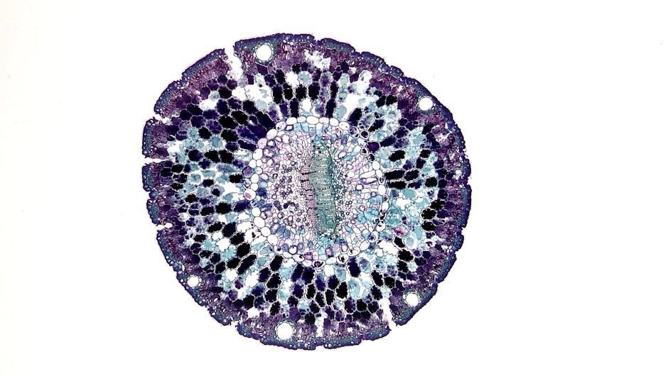
7.1 Anatomical Traits of Roots in Cereal Crops
Roots in cereal crops exhibit unique anatomical traits that enhance water and nutrient absorption. The epidermis‚ cortex‚ and stele are key layers‚ with the cortex often containing aerenchyma for aeration. Xylem and phloem tissues facilitate nutrient transport‚ while root hairs increase surface area for absorption. These structures are adapted to optimize soil resource utilization‚ ensuring plant growth and productivity. Research focuses on genetic and environmental factors influencing root architecture‚ aiming to improve crop resilience and yield in diverse agricultural conditions.
7.2 Research on Root Architecture and Function
Research on root architecture focuses on understanding the structural and functional diversity of roots in cereal crops; Advanced imaging techniques and genetic studies reveal how root traits influence nutrient uptake and water acquisition. Scientists investigate root plasticity‚ exploring how environmental factors shape root development. This research aims to enhance crop resilience and yield under varying conditions. By identifying key genetic factors‚ breeding programs can develop crops with optimized root systems‚ improving agricultural productivity and sustainability in challenging environments.
7.3 Genetic Factors Influencing Root Growth
Genetic factors play a crucial role in determining root growth patterns and architecture. Specific genes regulate cell elongation‚ division‚ and differentiation in root tissues. Mutagenesis and RNA interference studies have identified key genes involved in root development. Environmental interactions with genetic makeup shape root traits‚ influencing nutrient uptake efficiency. Understanding these genetic mechanisms allows for targeted breeding to enhance root systems in crops‚ improving drought tolerance and nutrient acquisition. This research bridges genetics and agriculture‚ promoting sustainable crop production.
Laboratory Data Collection and Analysis
Laboratory data collection involves systematic gathering of experimental information using specialized tools and techniques. Accurate recording and analysis of data are crucial for valid scientific conclusions.
8.1 Organizing and Interpreting Experimental Data
Organizing experimental data involves categorizing and structuring information for clarity. Proper interpretation requires understanding patterns and trends‚ ensuring accurate conclusions. Effective use of graphs and charts enhances visualization‚ while statistical methods validate findings. Clear documentation and ethical reporting are essential to maintain integrity and reproducibility in scientific research. This process equips students with skills to translate raw data into meaningful insights‚ fostering a deeper understanding of anatomical and physiological principles.
8.2 Statistical Analysis of Physiological Data
Statistical analysis is crucial for validating physiological data‚ ensuring accuracy and reliability. Common methods include descriptive statistics and inferential tests to identify trends and correlations. Tools like Excel or specialized software facilitate data processing. Understanding biological variability and experimental errors is essential for precise interpretation. Proper statistical techniques enhance the credibility of laboratory findings‚ aiding in informed decision-making and advancing research in anatomy and physiology. Accurate analysis ensures data integrity and supports meaningful conclusions in scientific studies and healthcare applications.
8.3 Reporting Findings in a Scientific Format
Reporting findings in a scientific format requires clear and concise presentation of data‚ analysis‚ and conclusions. Proper documentation includes titles‚ introductions‚ materials and methods‚ results‚ and discussions. Graphs‚ tables‚ and images should be labeled and referenced appropriately. Ensure accuracy in data representation and avoid bias in interpretations. Use standard scientific terminology and maintain objectivity. Adherence to formatting guidelines ensures consistency and professionalism‚ making findings accessible and understandable to peers and researchers in anatomy and physiology studies. Ethical reporting practices are essential for credibility.
Modern Tools in Anatomy and Physiology Education
Modern tools like simulation software and 3D printing enhance anatomy and physiology education‚ offering interactive and immersive learning experiences. These technologies provide detailed visualizations and hands-on practice‚ improving understanding of complex structures and functions. Online resources and virtual labs further support student engagement and accessibility‚ making learning more effective and enjoyable. These innovations are transforming traditional educational methods‚ fostering deeper comprehension and practical skills in anatomy and physiology.
9.1 Use of Simulation Software for Virtual Dissections
Simulation software revolutionizes anatomy and physiology education by offering virtual dissection experiences. These tools provide detailed‚ interactive 3D models‚ allowing students to explore complex structures from multiple angles. Virtual labs enhance engagement and accessibility‚ enabling repetitive practice without physical specimens. Simulation software also supports diverse learning styles‚ offering visual‚ tactile‚ and kinesthetic experiences. This technology complements traditional methods‚ fostering a deeper understanding of anatomical relationships and physiological processes. It prepares students for real-world scenarios‚ making it an invaluable resource in modern education.
9.2 3D Printing in Anatomy Education
3D printing is transforming anatomy education by creating detailed‚ customizable models of anatomical structures. These models allow students to explore complex tissues and organs in a tactile manner‚ enhancing spatial understanding. Printing patient-specific or rare anatomical variations enables hands-on study of unique cases. This technology fosters engagement and retention‚ making intricate structures more accessible. As costs decrease‚ 3D printing is becoming a valuable tool for institutions‚ offering a modern approach to anatomy training and supplementing traditional teaching methods effectively.
9.3 Online Resources for Anatomy and Physiology Learning
Online resources revolutionize anatomy and physiology education by providing interactive tools and accessible content. Platforms offer detailed diagrams‚ virtual dissections‚ and 3D models‚ enhancing visual learning. Websites like AnatomyTOOL and Visible Body provide immersive experiences‚ while apps such as Complete Anatomy enable students to explore complex structures on mobile devices. These resources support self-paced learning‚ allowing students to reinforce concepts outside the lab. Interactive simulations and quizzes further aid in understanding physiological processes‚ making online resources invaluable for modern anatomy education.

Assessment and Feedback in Laboratory Work
Assessment and feedback in lab work involve evaluating student performance through graded assignments‚ lab reports‚ and presentations. Constructive feedback helps refine skills and understanding.
10.1 Grading Criteria for Lab Reports and Presentations
Lab reports and presentations are graded based on clarity‚ accuracy‚ and adherence to scientific standards. Reports must include objectives‚ methods‚ results‚ and conclusions. Presentations require clear communication and visual aids. Proper citation and referencing are essential. Graders evaluate understanding‚ critical thinking‚ and the ability to interpret data. Peer reviews and collaborative feedback enhance learning and improvement. Consistent effort and professionalism in submissions significantly impact final grades. Attention to detail ensures comprehensive assessment of student performance.
10.2 Peer Review and Collaborative Learning
Peer review and collaborative learning enhance the educational experience by fostering critical thinking and communication skills. Students review each other’s work‚ providing constructive feedback to improve understanding and accuracy. This interactive process encourages active participation and mutual learning. Collaborative activities promote teamwork‚ shared knowledge‚ and problem-solving. Feedback from peers helps identify strengths and weaknesses‚ while group discussions refine ideas and concepts. This approach prepares students for professional environments where teamwork and clear communication are essential. It also builds confidence and interpersonal skills.
10.3 Improving Lab Performance Based on Feedback
Feedback is a vital tool for enhancing lab performance and fostering growth. By analyzing instructor and peer reviews‚ students can identify areas for improvement and refine their techniques. Implementing feedback leads to better experimental design‚ improved data collection‚ and enhanced scientific reasoning; Regular reflection on performance helps develop a systematic approach to problem-solving and strengthens understanding of anatomical and physiological concepts. This iterative process cultivates resilience and adaptability‚ essential for success in scientific and professional environments. Continuous improvement ensures mastery of lab skills and intellectual growth.
Safety Protocols in the Lab
Lab safety is crucial to protect students and equipment. Proper handling of chemicals‚ biological specimens‚ and sharp tools is emphasized. Emergency procedures‚ fire drills‚ and spill management are regularly practiced. Personal protective equipment (PPE) is mandatory‚ and waste disposal guidelines are strictly followed. Adhering to these protocols ensures a safe and responsible learning environment for all participants. Regular training and updates keep everyone informed and prepared for potential hazards.
11;1 Handling Chemicals and Biological Specimens Safely
Proper handling of chemicals and biological specimens is essential to prevent accidents and exposure. Always wear personal protective equipment (PPE)‚ including gloves‚ goggles‚ and lab coats. Chemicals should be stored in labeled containers and used in well-ventilated areas. Biological specimens must be handled with care to avoid contamination. Dispose of hazardous waste according to guidelines‚ and follow safety data sheets (SDS) for specific substances. Spills should be contained and cleaned immediately‚ and emergency equipment like eyewashes and fire extinguishers should be accessible. Regular training and supervision ensure compliance with safety protocols.
11.2 Emergency Procedures in the Lab
In case of emergencies‚ evacuate the area immediately and alert others. Use fire extinguishers for small fires and know the location of emergency exits. For chemical spills‚ contain the area and use absorbent materials. In case of exposure‚ flush with water or wash skin thoroughly. For biological specimen incidents‚ disinfect and seek medical help if necessary. Always activate alarms and contact emergency services when needed. Post-emergency‚ document incidents and receive first aid if required. Regular drills ensure preparedness and quick response.
11.3 Waste Disposal and Environmental Responsibility
Proper waste disposal is crucial for maintaining a safe and environmentally responsible lab environment. Separate hazardous and non-hazardous waste‚ using designated containers. Chemical waste must be labeled and stored according to regulations. Biological specimens and materials should be disposed of in biohazard bins. Recyclable items should be segregated to minimize environmental impact. Personnel must wear protective gear when handling waste. Regular training on waste management ensures compliance with safety and environmental standards. Proper disposal practices protect both human health and the planet.