
X-ray positioning charts are visual guides that illustrate optimal patient positioning for specific radiographic projections. They include anatomical landmarks, positioning guidelines, and reference images for accurate imaging.
1.1 Definition and Purpose
An X-ray positioning chart is a detailed visual guide that outlines the optimal positioning for patients during radiographic examinations. It serves as a standardized tool for radiographers to ensure accurate and diagnostic image quality. The primary purpose of these charts is to provide clear, step-by-step instructions for aligning patients correctly, minimizing retakes, and ensuring patient safety. They are designed to accommodate various anatomical structures and pathological conditions, offering a consistent approach to imaging. By following the chart, radiographers can achieve precise alignment of the X-ray beam with the target anatomy, reducing errors and enhancing diagnostic accuracy. These charts are essential for maintaining quality and consistency in radiographic procedures across different clinical settings.
1.2 Importance in Radiography
X-ray positioning charts are fundamental in radiography as they ensure consistent, high-quality imaging. They guide radiographers in achieving precise patient alignment, reducing errors and retakes. By standardizing positioning, these charts improve diagnostic accuracy and enhance patient safety. They also help minimize radiation exposure by ensuring the X-ray beam is correctly focused. Additionally, the charts act as educational tools, aiding students and professionals in understanding proper techniques. Their use promotes efficiency in clinical workflows, allowing for faster and more accurate image acquisition. Overall, X-ray positioning charts are essential for maintaining high standards in radiographic imaging, ensuring reliable and reproducible results across diverse clinical settings.
1.3 Overview of the Chart Structure
An X-ray positioning chart is a structured guide that outlines the optimal setup for radiographic examinations. It typically includes detailed anatomical landmarks, patient positioning instructions, and technical parameters such as exposure settings. The chart often features reference images to illustrate proper alignment and expected outcomes. These visual aids help ensure consistency and accuracy in imaging. The structure is organized by body regions or specific projections, making it easy to navigate. Charts may also incorporate symbols or annotations to highlight key positioning points. By following the chart, radiographers can achieve high-quality images that meet diagnostic standards. This standardized approach minimizes variability and enhances the reliability of radiographic results.

Key Components of an X-Ray Positioning Chart
X-ray positioning charts include anatomical landmarks, patient positioning guidelines, equipment setup, and reference images for accurate radiographic projections, ensuring proper alignment and diagnostic-quality results.
2.1 Anatomical Landmarks
Anatomical landmarks are specific body structures used to guide accurate patient positioning during X-ray imaging. These landmarks, such as the clavicle for chest X-rays or the iliac crest for abdominal imaging, help radiographers align the X-ray beam correctly. They ensure proper positioning, reducing the need for retakes and improving diagnostic quality. Landmarks vary by body region and projection, requiring precise identification to achieve optimal results. For example, the spinous processes are often used for spinal imaging, while the malleoli are key for ankle X-rays. These reference points are visually represented in positioning charts, making them essential for consistent and accurate radiographic outcomes. Their use ensures standardization and reliability in imaging procedures across different patient populations and anatomical regions.
2.2 Patient Positioning Guidelines
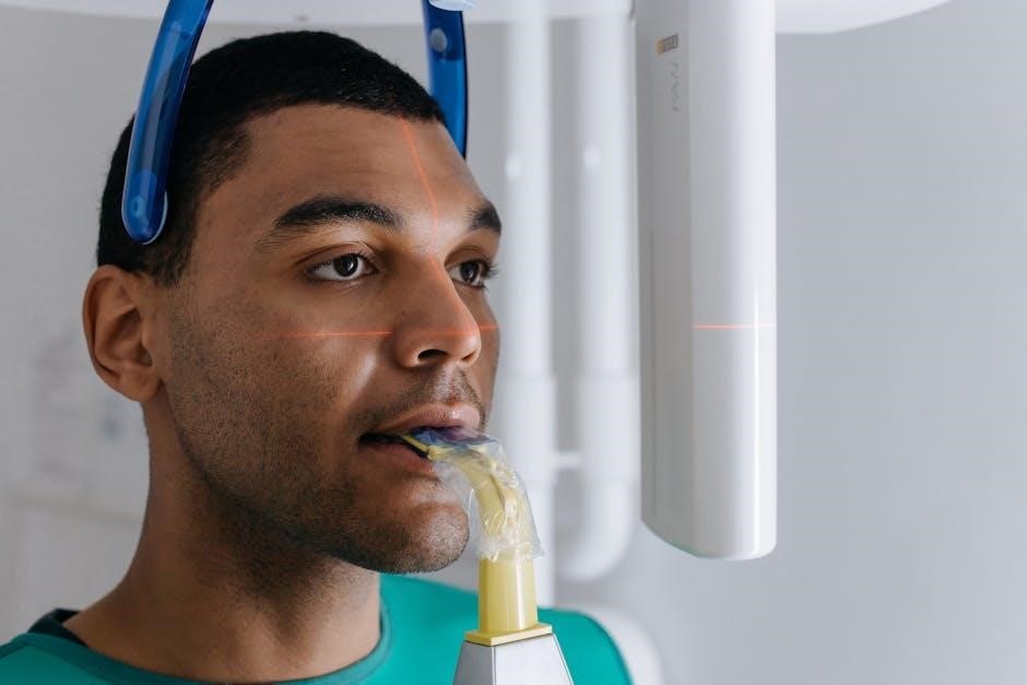
Patient positioning guidelines outline the specific poses and alignments required for accurate X-ray imaging. These guidelines ensure the correct placement of body parts relative to the X-ray beam, minimizing distortions and ensuring diagnostic clarity. Proper positioning involves aligning the patient’s body with the image receptor and adjusting posture to achieve the desired view. For example, chest X-rays require the patient to stand upright with hands on hips, while spinal X-rays may involve lying down with knees slightly flexed. Movement restriction, such as holding breath, is often necessary to prevent blurring. These guidelines are standardized to ensure consistency and accuracy across imaging procedures. Adherence to positioning guidelines is critical for obtaining high-quality images and ensuring patient safety during radiographic examinations.
2.3 Equipment Setup and Alignment
Proper equipment setup and alignment are crucial for obtaining accurate X-ray images. The X-ray table and grid must be positioned correctly to ensure optimal image quality. The X-ray beam should be aligned with the anatomical landmarks specified in the positioning chart, and the central ray must be perpendicular to the image receptor. Adjustments to the table height, tube angle, and source-to-image receptor distance (SID) are often necessary to accommodate patient size and the desired projection. Alignment markers and laser guides on modern X-ray machines help in precise positioning; Ensuring the equipment is properly calibrated and aligned minimizes artifacts and enhances diagnostic clarity. Regular maintenance of the X-ray unit and accessories is also essential to maintain consistent performance and image quality.
2.4 Reference Images for Comparison
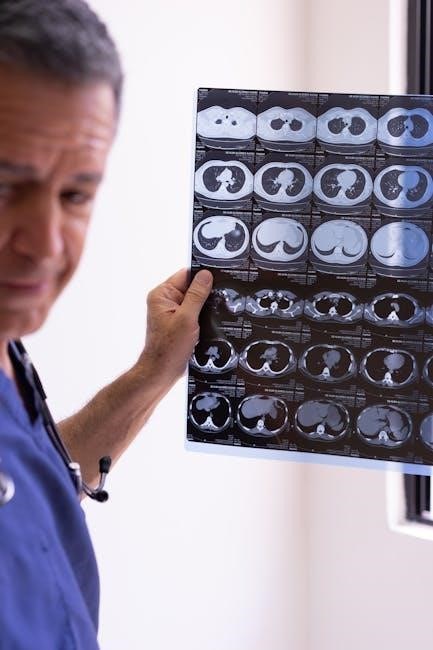
Reference images are essential components of X-ray positioning charts, providing visual comparisons for accurate radiographic results. These images, often included in PDF formats, depict ideal anatomical alignment and tissue representation for specific projections. By comparing patient images to these references, radiographers can identify deviations and adjust positioning to improve diagnostic accuracy. Reference images also help in recognizing abnormalities or artifacts, ensuring consistent and reliable outcomes. They are particularly useful for training purposes, allowing professionals to familiarize themselves with normal and abnormal anatomical variations. The inclusion of these images in positioning charts enhances the learning process and supports precise patient positioning, ultimately contributing to higher-quality X-ray examinations and better patient care.
How to Use an X-Ray Positioning Chart
X-ray positioning charts guide radiographers in aligning patients correctly for precise imaging. They outline steps for preparing the patient, positioning, and adjusting equipment for optimal results.
3.1 Preparing the Patient
Preparing the patient involves ensuring they are comfortable and properly positioned. Remove any obstructions like jewelry or clothing that may interfere with the X-ray beam. Explain the procedure to reduce anxiety and ensure cooperation. Position the patient according to the chart’s guidelines, using anatomical landmarks as references. Ensure the body part to be imaged is accessible and aligned correctly. Adjust the patient’s posture to maintain natural positioning unless specified otherwise. Use supportive devices if necessary to stabilize the patient during the examination. Verify exposure parameters like kVp and mAs to ensure optimal image quality. This step is crucial for obtaining clear, diagnostic images and minimizing retakes.
3.2 Aligning the X-Ray Beam
Aligning the X-ray beam correctly is essential for obtaining clear, diagnostic images. Use the positioning chart to guide the beam’s center and angle, ensuring it corresponds to the anatomical landmarks. Reference images in the chart help verify proper alignment. Adjust the beam’s height, angle, and rotation to match the patient’s position, as indicated in the chart. Ensure the beam is centered on the body part being imaged and that the grid or film is properly aligned. Correct alignment minimizes artifacts and ensures optimal image quality. This step is critical for accurate diagnostics and patient safety, as misalignment can lead to retakes or incomplete information.
3.3 Adjusting Positioning for Optimal Results
Adjusting patient positioning is crucial to ensure diagnostic-quality images. Use the X-ray positioning chart as a guide to fine-tune the patient’s alignment. Reference images in the chart help identify and correct misalignments. Pay attention to anatomical landmarks and adjust the patient’s posture, rotation, or angle as needed. Ensure the X-ray beam is properly aligned with the target anatomy to avoid distortion or artifacts. For optimal results, consider the patient’s body size, shape, and any physical limitations. Iterative adjustments may be necessary to achieve the desired positioning. Regularly compare the setup with the chart to ensure accuracy and consistency. Proper adjustments enhance image clarity, reducing the need for retakes and improving diagnostic outcomes.

Common X-Ray Projections and Their Positioning
X-ray positioning charts detail standard projections like chest (PA/AP), spine (AP/lateral), limb (upper/lower extremities), and skull (AP/lateral) views. Each projection has specific anatomical alignment requirements.
4.1 Chest X-Ray (PA and AP Views)
The chest X-ray is a fundamental radiographic projection, commonly performed in posteroanterior (PA) and anteroposterior (AP) views. The PA view, taken with the patient standing, provides a clear image of the lungs, heart, and mediastinum. The AP view is often used for bedridden or non-ambulatory patients, offering similar diagnostic value. Proper patient positioning is critical: for PA, the patient stands upright, hands on hips, and chest against the image receptor. For AP, the patient lies supine, with arms extended overhead. Both views require the X-ray beam to be centered at the midthoracic spine. These projections are essential for diagnosing respiratory and cardiac conditions, ensuring accurate anatomical representation. Proper alignment and breathing instructions are vital for optimal image quality and diagnostic accuracy.
4.2 Spinal X-Ray (AP and Lateral Views)
Spinal X-rays are essential for evaluating vertebral alignment, disc integrity, and spinal pathologies. The anteroposterior (AP) view captures the spine in a frontal plane, requiring the patient to stand or lie supine with the X-ray beam centered at the midthoracic spine. The lateral view provides a side profile, typically taken with the patient standing or sitting upright, ensuring the beam is aligned with the midsagittal plane. Proper positioning involves ensuring the vertebral bodies and spinous processes are clearly visible. For both views, anatomical landmarks such as the C7 vertebra or iliac crests are used for reference. These projections are critical for diagnosing conditions like fractures, scoliosis, or spondylolisthesis. Correct patient alignment and breathing techniques are vital for obtaining clear, diagnostic images.
4.3 Limb X-Ray (Upper and Lower Extremities)
Limb X-rays are used to evaluate bones, joints, and soft tissues in the arms and legs. For upper extremities, such as the humerus or forearm, the patient is positioned with the limb extended and aligned with the image receptor. Lower extremity X-rays, like femur or tibia/fibula views, require the patient to stand or lie supine with the limb straight. Proper alignment ensures the X-ray beam is perpendicular to the area of interest. Reference images in positioning charts help technicians achieve accurate and consistent results. These projections are vital for diagnosing fractures, joint dislocations, or bone abnormalities. Correct patient positioning and beam alignment are critical to obtaining clear, diagnostic images of upper and lower limbs.
4.4 Skull X-Ray (AP and Lateral Views)
Skull X-rays are performed to evaluate cranial structures, including fractures, abnormalities, or pathologies. The anteroposterior (AP) view requires the patient to be upright or supine, with the skull centered and the X-ray beam angled to avoid distortion. The lateral view involves the patient sitting or standing with the head positioned against the image receptor, ensuring the petrous ridge is visible. Proper alignment is critical to capture clear images of the cranial vault and facial bones. Positioning charts provide detailed guidelines for beam angulation and patient placement. Reference images help ensure accurate and consistent results, making these projections essential for diagnosing skull-related conditions. Proper technique minimizes artifacts and ensures diagnostic clarity in both AP and lateral skull X-rays.

Best Practices for X-Ray Positioning
Adhering to standardized positioning guidelines ensures consistency and accuracy. Using reference images and clear patient communication enhances diagnostic quality. Continuous learning improves technique and safety.
5.1 Adherence to Standard Guidelines
Adhering to standardized guidelines in X-ray positioning ensures consistency and accuracy across all radiographic procedures. These guidelines provide detailed instructions for patient positioning, equipment alignment, and exposure parameters. They are developed based on evidence-based practices and expert recommendations to optimize image quality. By following these standards, radiographers can minimize errors and ensure reproducibility of images. Proper patient positioning is critical for obtaining diagnostic-quality images, as misalignment can lead to artifacts or incomplete visualization of anatomical structures. Standard guidelines also help in reducing radiation exposure by ensuring that the X-ray beam is correctly collimated and aligned. This adherence promotes patient safety, improves diagnostic accuracy, and supports efficient workflow in radiography departments. Regular updates to these guidelines reflect advancements in technology and techniques, ensuring they remain relevant and effective.
5.2 Patient Communication and Safety
Patient communication and safety are paramount in radiographic procedures. Clear communication helps reduce anxiety and ensures patient cooperation during positioning. Radiographers must explain the procedure, positioning requirements, and any necessary breathing instructions. Ensuring patient safety involves minimizing radiation exposure by using appropriate shielding and collimation. Positioning charts often include safety protocols to guide radiographers in protecting sensitive areas. Proper alignment and immobilization techniques prevent movement artifacts and ensure accurate imaging. Open dialogue fosters trust and compliance, leading to better diagnostic outcomes. Safety measures also include verifying patient identity and medical history to avoid contraindications. By prioritizing communication and safety, radiographers create a positive experience while maintaining high standards of care and image quality.
5.3 Continuous Learning and Updates
Continuous learning and updates are essential for radiographers to stay current with advancements in X-ray positioning techniques. Regular training ensures familiarity with new equipment and positioning charts, improving image quality and diagnostic accuracy. Radiographers should engage in workshops, webinars, and professional courses to refine their skills. Staying updated with industry standards and evidence-based practices is crucial for delivering optimal patient care. Digital positioning charts and integrated imaging systems require ongoing education to maximize their benefits. Peer reviews and feedback sessions also foster professional growth. By committing to lifelong learning, radiographers adapt to technological advancements and evolving patient needs, ensuring precise and safe positioning for accurate radiographic outcomes.
Troubleshooting Common Positioning Errors
Troubleshooting positioning errors involves identifying misalignment, artifacts, and improper exposures. Adjustments are made to optimize image quality, ensuring accurate diagnostics and minimizing retakes.
6.1 Identifying Misalignment
Identifying misalignment in X-ray positioning involves analyzing the anatomical landmarks and their alignment with the X-ray beam. Visual cues such as uneven density, distorted anatomy, or rotation can indicate misalignment. Referencing positioning charts helps compare expected and actual alignments. For instance, in chest X-rays, misalignment may result in uneven lung fields or off-center mediastinum. Proper use of reference images from positioning charts aids in detecting such errors. Adjustments are made by repositioning the patient or the X-ray beam to ensure accurate and diagnostically useful images. Regular training and adherence to standardized guidelines minimize misalignment, ensuring optimal radiographic outcomes and reducing the need for retakes. Continuous learning and updates in positioning techniques further enhance accuracy.
6.2 Adjusting for Artifacts in Images
Artifacts in X-ray images, such as streaks, blur, or distortion, can compromise diagnostic quality. These artifacts often arise from improper positioning, movement, or equipment misalignment. Adjusting for artifacts involves reviewing reference images from positioning charts to identify deviations. For instance, grid cutoff or patient motion artifacts can be corrected by repositioning the patient or adjusting the X-ray beam angle. Additionally, ensuring proper alignment of anatomical landmarks with the central ray helps minimize artifacts. Using positioning charts with annotated examples allows for precise adjustments, ensuring clearer and more accurate images. Regular training and adherence to standardized protocols are essential for recognizing and correcting artifacts effectively, improving overall image quality and diagnostic confidence. Continuous learning and updates in positioning techniques further enhance the ability to identify and address artifacts promptly.
Role of Technology in X-Ray Positioning
Technology enhances X-ray positioning accuracy and efficiency through digital charts, AI-driven software, and real-time adjustments. These tools reduce errors and improve image quality for better diagnostics.
7.1 Digital Positioning Charts
Digital positioning charts have revolutionized radiography by providing interactive and customizable guides for X-ray positioning. These charts are accessible via PDFs and online platforms, offering detailed anatomical landmarks, exposure parameters, and reference images. They allow radiographers to quickly access and compare positioning setups, reducing errors and improving efficiency; Digital charts often include interactive features, such as zoom functionality and searchable databases, making them more user-friendly. Additionally, they can be integrated with imaging systems, enabling real-time adjustments during procedures. This technology ensures consistency and accuracy, especially in complex or specialized cases. By leveraging digital tools, radiographers can enhance patient safety and diagnostic outcomes, staying updated with the latest imaging techniques and guidelines.
7.2 Integration with Imaging Systems
Digital positioning charts can seamlessly integrate with modern imaging systems, enhancing workflow efficiency and accuracy. This integration allows radiographers to access positioning guidelines directly on the X-ray system’s interface, ensuring consistency and reducing errors. Imaging systems can automatically adjust exposure parameters based on the chart’s recommendations, optimizing image quality. Additionally, integrated systems provide real-time feedback, enabling immediate adjustments for better diagnostic outcomes. This technology also supports the storage and retrieval of patient-specific positioning data, streamlining future procedures. By combining digital charts with imaging systems, radiographers can achieve precise and reproducible results, improving patient care and operational efficiency. This integration is a cornerstone of modern radiography, enabling faster and more accurate imaging processes.

X-Ray Positioning Charts for Specialized Cases
X-ray positioning charts for specialized cases, such as pediatric or geriatric patients, provide tailored guidelines to accommodate unique anatomical and positioning requirements, ensuring accurate and safe imaging.
8.1 Pediatric Patients
Pediatric patients require specialized X-ray positioning due to their smaller body size and developing anatomy. Positioning charts for children often include scaled-down guidelines and adjusted anatomical landmarks to accommodate growth stages. Techniques may involve smaller X-ray fields, lower radiation doses, and additional support devices to ensure proper alignment and comfort. The charts frequently incorporate reference images and detailed instructions to help radiographers achieve accurate and safe imaging for young patients; These adaptations ensure that the unique needs of pediatric cases are met, providing clear and diagnostic images while minimizing radiation exposure. The use of tailored charts helps in reducing errors and improving outcomes in pediatric radiography.
8.2 Geriatric Patients
Geriatric patients often require specialized X-ray positioning due to mobility issues, fragile bones, and potential spinal degeneration. Positioning charts for elderly patients emphasize comfort and safety, with adjustments for limited movement or physical disabilities. Supportive devices, such as cushions or braces, are frequently recommended to maintain proper alignment and reduce discomfort. Clear communication is crucial to ensure patient cooperation, as anxiety or pain may hinder accurate positioning. Radiation safety is prioritized, with techniques optimized to minimize exposure while achieving diagnostic-quality images. The charts also include reference images to account for age-related anatomical changes, ensuring accurate and effective imaging for this vulnerable population. These tailored approaches help radiographers address the unique challenges of geriatric radiography.
X-ray positioning charts are essential for accurate radiographic imaging, ensuring proper alignment and minimizing radiation exposure. They provide clear guidelines and reference images for optimal results in diagnostics.
9.1 Summary of Key Points
X-ray positioning charts are visual guides that help radiographers accurately position patients for specific radiographic projections. They include anatomical landmarks, patient positioning guidelines, and reference images to ensure clear and accurate imaging. These charts minimize radiation exposure and enhance diagnostic quality by providing standardized procedures. They are essential for maintaining consistency in radiographic techniques and are widely used in medical imaging practices. The charts cover various projections, such as chest, spinal, and limb X-rays, offering a comprehensive resource for radiographers. By following these guidelines, professionals can achieve optimal results and improve patient outcomes. The use of positioning charts also supports continuous learning and adherence to safety protocols in radiography.
9.2 Future Trends in X-Ray Positioning
Future advancements in X-ray positioning are expected to integrate digital and interactive charts, enhancing precision and accessibility. Digital positioning charts with real-time adjustments and AI-driven recommendations will likely become standard, reducing errors and improving imaging outcomes. Integration with advanced imaging systems will enable seamless data sharing and enhanced diagnostic capabilities. The development of adaptive charts for specialized cases, such as pediatric and geriatric patients, will further personalize radiographic procedures. Additionally, the incorporation of 3D imaging and augmented reality tools may revolutionize how positioning is planned and executed. These innovations aim to streamline workflows, reduce radiation exposure, and improve patient care. The evolution of positioning charts will continue to play a pivotal role in advancing radiography.