
What is a Sleep Deprivation EEG? This diagnostic test enhances seizure detection‚ particularly for idiopathic generalized epilepsy‚ by intentionally inducing sleepiness before recording brain activity;
Why Perform an EEG After Sleep Deprivation? Sleep deprivation increases the likelihood of detecting epileptiform abnormalities‚ improving diagnostic sensitivity‚ though specificity requires careful consideration due to potential false positives.
What is a Sleep Deprivation EEG?
A Sleep Deprivation EEG (SD-EEG) is a neurophysiological test used to evaluate brain activity after a period of intentionally reduced sleep. It’s an extension of a standard Electroencephalogram (EEG)‚ but specifically designed to increase the chances of detecting subtle seizure patterns that might not be apparent during a normal waking state. The core principle relies on the fact that sleep deprivation can lower the seizure threshold in individuals predisposed to epilepsy.
During an SD-EEG‚ electrodes are attached to the scalp to record electrical impulses generated by the brain. The test aims to provoke epileptiform discharges – abnormal brain activity indicative of a seizure focus – making diagnosis more accurate‚ especially in cases of suspected idiopathic generalized epilepsy. It’s a sensitive tool‚ but interpretation requires careful consideration of potential false positives‚ as even healthy individuals can exhibit changes after sleep loss.
Why Perform an EEG After Sleep Deprivation?
An EEG performed after sleep deprivation is undertaken to enhance diagnostic sensitivity‚ particularly when investigating idiopathic generalized epilepsy. Standard EEGs may yield normal results even in individuals with a predisposition to seizures; sleep deprivation lowers the seizure threshold‚ increasing the probability of capturing epileptiform activity. This technique serves as a “trigger maneuver‚” attempting to provoke abnormal brainwave patterns that would otherwise remain undetected.
However‚ it’s crucial to acknowledge that SD-EEG isn’t foolproof. Specificity is a concern‚ as up to 12% of healthy individuals may exhibit interictal epileptiform activity (IEA) post-deprivation. Therefore‚ results must be interpreted cautiously‚ considering the patient’s clinical context and ruling out other potential causes for observed abnormalities.
Preparing for a Sleep Deprivation EEG
Understanding the Goal of Sleep Deprivation Maintaining wakefulness before the EEG aims to amplify abnormal brain activity‚ aiding in accurate seizure detection and diagnosis.
Age-Specific Considerations for Sleep Deprivation Protocols vary; CHOC schedules tests around nap times for infants‚ requiring parental patience and understanding.
Understanding the Goal of Sleep Deprivation
The primary objective of sleep deprivation prior to an EEG is to heighten the brain’s excitability‚ thereby increasing the probability of capturing epileptiform discharges that might otherwise remain undetected during a standard EEG recording. Normal sleep patterns often mask underlying seizure activity‚ and intentionally disrupting these patterns can unmask these abnormalities.
This technique is particularly valuable in diagnosing idiopathic generalized epilepsy‚ where seizure origins are widespread. By inducing a state of sleepiness‚ the brain becomes more susceptible to exhibiting these characteristic patterns. However‚ it’s crucial to remember that sleep deprivation isn’t a guaranteed solution; epileptiform changes can also occur in healthy individuals‚ necessitating careful interpretation of results. The goal isn’t simply to induce fatigue‚ but to strategically alter brain states for improved diagnostic accuracy.
Age-Specific Considerations for Sleep Deprivation
Sleep deprivation protocols must be tailored to the patient’s age. Infants and Toddlers require careful scheduling to coincide with nap times‚ acknowledging the difficulty of prolonged wakefulness and the emotional distress it causes. Children may require more creative strategies to maintain wakefulness‚ balancing the need for sleep loss with their well-being.
Adults generally tolerate longer periods of sleep deprivation‚ but cognitive impairment and irritability must be monitored. Maintaining wakefulness is challenging across all ages‚ demanding parental/guardian involvement and understanding. CHOC Children’s emphasizes patience and support‚ recognizing the hardship for both the child and their family throughout the process.
Infants and Toddlers
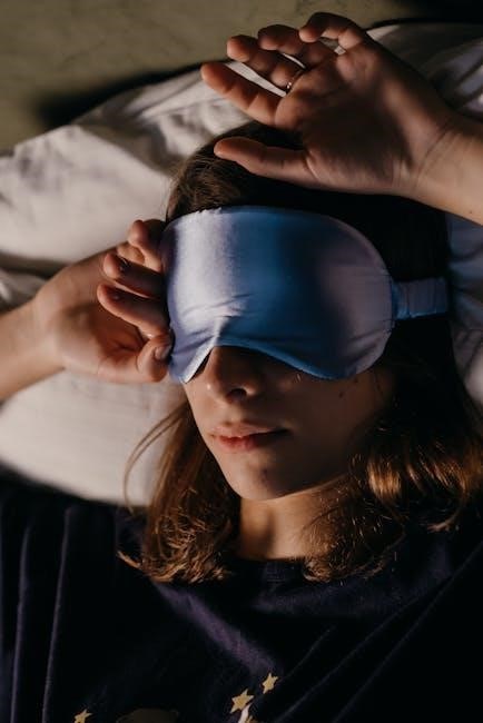
For infants and toddlers undergoing sleep deprivation EEG‚ the process necessitates a delicate approach. CHOC Children’s prioritizes scheduling the test to align with the baby’s natural nap schedule‚ recognizing the inherent difficulty in keeping them awake for extended periods. Maintaining wakefulness in this age group is particularly challenging and can be emotionally taxing for both the child and parents.
Caregivers should focus on minimizing distress and maximizing comfort. Brief‚ engaging activities can help‚ but prolonged attempts to force wakefulness are discouraged. The goal is to gently disrupt the sleep cycle‚ not to cause significant upset. Patience and understanding are paramount throughout the preparation and recording.
Children
Preparing children for a sleep deprivation EEG requires a balance of maintaining wakefulness and minimizing frustration. As with infants‚ CHOC emphasizes the difficulty for both the child and parents. Engaging activities‚ such as games or reading‚ can help keep older children awake during the designated period before the test.
However‚ it’s crucial to avoid overstimulation‚ which could interfere with the EEG recording. Parental patience and a calm demeanor are essential. Explaining the process in age-appropriate terms can also reduce anxiety. Remember‚ the aim is gentle sleep cycle disruption‚ not prolonged distress or forced wakefulness.
Adults
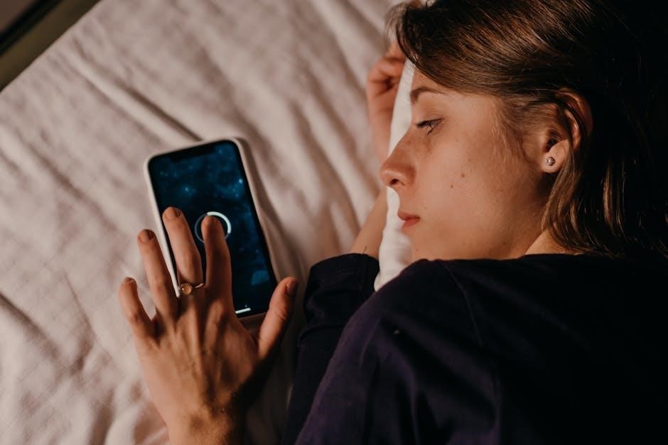
For adult patients undergoing a sleep deprivation EEG‚ maintaining wakefulness typically involves restricting nighttime sleep. Avoid daytime naps and engage in stimulating activities. However‚ it’s vital to prioritize safety; avoid driving or operating heavy machinery during the sleep-deprived state.
Unlike children‚ adults can often understand the rationale behind the protocol‚ facilitating cooperation. Clear communication regarding the importance of adhering to the sleep schedule is key. While caffeine might seem helpful‚ discuss its use with the medical team‚ as it can potentially influence EEG results. Prioritize a calm and quiet environment before the test.
Parental/Guardian Role in Maintaining Wakefulness
Parents or guardians play a crucial role in ensuring the child remains awake before the EEG. This requires a dedicated effort‚ understanding it can be challenging and emotionally taxing for both the child and themselves. Distraction with engaging activities – games‚ stories‚ or gentle play – is highly effective.
Avoid screen time close to bedtime‚ as the blue light can interfere with sleepiness. Patience and understanding are paramount‚ acknowledging the child’s increasing irritability. CHOC recommends scheduling the test to coincide with nap times for infants‚ but consistent wakefulness is the goal. Remember‚ a cooperative child yields better results.
Medication Considerations Before the EEG
It’s vital to inform the neurologist of all medications the child is taking‚ as some can influence EEG results. Certain medications may suppress seizure activity‚ potentially masking abnormalities‚ or conversely‚ lower the seizure threshold. Discuss with the doctor whether any medications should be temporarily adjusted or held before the test.
Specifically‚ inquire about anti-epileptic drugs and any medications affecting sleep or alertness. The use of melatonin to aid sleep during the EEG recording is sometimes proposed‚ but its impact on the sleep deprivation process itself needs careful consideration and physician approval. Always follow the neurologist’s specific instructions regarding medication management.
The EEG Procedure Itself
Standard EEG Setup involves electrode placement on the scalp using a conductive gel. Modifications for Sleep Deprivation EEG may include net electrodes for improved signal quality.
Duration of the Sleep Deprivation EEG Recording typically lasts at least 2-3 hours‚ with maximum effort to obtain a sleep record during the test.
Standard EEG Setup
The conventional EEG setup begins with meticulous preparation of the scalp‚ ensuring it’s clean and free from oils or hair products that could impede signal transmission. A cap containing numerous disc-shaped electrodes is then carefully applied to the patient’s head‚ utilizing a conductive gel to minimize impedance and maximize the quality of the recorded brainwaves.
These electrodes are strategically positioned according to a standardized system – typically the 10-20 system – to cover all regions of the cerebral cortex. Each electrode is connected via wires to an EEG amplifier‚ which detects and amplifies the incredibly faint electrical signals generated by neuronal activity. Throughout the recording‚ technicians continuously monitor electrode impedance to ensure optimal contact and signal clarity.
The entire process aims to create a reliable and consistent baseline for detecting any changes in brain activity‚ especially those induced by sleep deprivation.
Modifications for Sleep Deprivation EEG
While the fundamental EEG setup remains consistent‚ sleep deprivation EEG necessitates specific adjustments to optimize its diagnostic yield. Recording is often scheduled in the afternoon to capitalize on the patient’s naturally lower alertness and facilitate sleep onset. Utilizing net electrodes is also recommended for improved signal quality and comfort during extended monitoring.
Technicians prioritize maximizing efforts to obtain a sleep record‚ as the transition into sleep is when epileptiform discharges are most likely to emerge. The recording duration is extended to at least 2-3 hours‚ allowing ample time to capture these events. Furthermore‚ the potential use of melatonin is considered to aid sleep induction‚ particularly in individuals struggling to fall asleep.
Duration of the Sleep Deprivation EEG Recording
The length of a sleep deprivation EEG recording is a crucial factor influencing its effectiveness. Standard EEGs are typically shorter‚ but to maximize the detection of epileptiform activity following sleep deprivation‚ a prolonged recording period is essential. Current proposed guidelines suggest a minimum recording duration of 2-3 hours‚ though this can be adjusted based on individual patient factors and clinical presentation.
This extended timeframe allows technicians to capture the brain’s activity as the patient transitions through different sleep stages‚ increasing the probability of identifying subtle or intermittent seizure patterns. Continuous monitoring throughout this period is vital for comprehensive data analysis and accurate interpretation.

Interpreting the Results
Normal EEG Findings After Sleep Deprivation Up to 12% of healthy individuals may exhibit interictal epileptiform activity (IEA) post-deprivation‚ necessitating careful differentiation from pathological findings.
Normal EEG Findings After Sleep Deprivation
Evaluating a sleep-deprived EEG requires acknowledging that epileptiform discharges can occur even in neurologically normal individuals. Studies indicate that a significant proportion – potentially up to 12% – of healthy subjects may demonstrate interictal epileptiform activity (IEA) following sleep deprivation.
Therefore‚ the presence of IEA alone doesn’t automatically confirm a diagnosis of epilepsy; context is crucial. The interpretation must consider the clinical picture‚ EEG background‚ and morphology of the observed discharges. Distinguishing these benign variations from true pathological activity is a key challenge.
Clinicians must carefully assess the EEG‚ looking for patterns consistent with normal sleep deprivation effects versus those indicative of an underlying epileptic disorder. A comprehensive analysis is essential for accurate diagnosis.
Interpreting Interictal Epileptiform Activity (IEA)
When interpreting IEA on a sleep-deprived EEG‚ careful consideration of several factors is paramount. The morphology‚ frequency‚ and distribution of spikes or sharp waves are crucial. Generalized IEA‚ appearing bilaterally and symmetrically‚ is more suggestive of idiopathic generalized epilepsy.
However‚ even focal IEA can be observed in healthy individuals post-deprivation. The EEG background activity must also be assessed; slowing or asymmetry can indicate underlying pathology. Correlation with the patient’s clinical history and neurological examination is essential.
Ultimately‚ IEA’s significance is determined by its context‚ not its mere presence‚ demanding a nuanced and comprehensive evaluation.
Sensitivity and Specificity of SD-EEG
The sensitivity of sleep deprivation EEG (SD-EEG) for detecting epilepsy‚ particularly idiopathic generalized epilepsy‚ remains a topic of ongoing debate. While SD-EEG can increase the yield of epileptiform abnormalities compared to standard EEG‚ its sensitivity isn’t absolute.
Specificity is a significant concern‚ as up to 12% of healthy individuals may exhibit interictal epileptiform activity (IEA) after sleep deprivation. This highlights the importance of interpreting findings cautiously and integrating them with clinical data.
Balancing sensitivity and specificity is crucial for accurate diagnosis‚ avoiding both false negatives and false positives.
Distinguishing Between Pathological and Benign Findings
Differentiating between pathological and benign findings on a sleep-deprived EEG requires careful consideration. The presence of interictal epileptiform discharges (IEDs) doesn’t automatically confirm epilepsy; they must be evaluated within the clinical context.
Factors like the morphology‚ frequency‚ and distribution of IEDs are crucial. The occurrence of IEDs in healthy individuals post-sleep deprivation necessitates a nuanced interpretation‚ avoiding overdiagnosis.
Correlation with the patient’s history‚ neurological examination‚ and other diagnostic tests is essential for accurate assessment and appropriate management decisions.

Sleep Deprivation Protocols & Standardization
Variability in Protocols Across Centers Currently‚ there’s no universally accepted standard for sleep deprivation EEG length or methodology‚ leading to inconsistencies between neurological centers.
Proposed Guidelines for Standardized Recording Recent proposals suggest using net electrodes‚ afternoon scheduling‚ and at least 2-3 hours of recording‚ potentially with melatonin assistance.
Variability in Protocols Across Centers
A significant challenge in interpreting sleep deprivation EEG (SD-EEG) results stems from the wide range of protocols employed across different neurological centers. Currently‚ a standardized approach is lacking‚ leading to considerable variability in how these tests are conducted. This inconsistency primarily revolves around the duration of sleep deprivation itself; there’s no defined standard length‚ with practices differing substantially between institutions.
Furthermore‚ variations exist in recording parameters and the specific techniques used during the EEG. Some centers may utilize different electrode placements or recording durations post-sleep deprivation. This lack of uniformity complicates comparisons between studies and can impact the reliability of diagnostic interpretations. Addressing this variability is crucial for enhancing the clinical utility of SD-EEG and ensuring consistent patient care.
Proposed Guidelines for Standardized Recording
Recognizing the current inconsistencies‚ efforts are underway to establish minimum standards for SD-EEG recording. Recent proposals advocate for utilizing net electrodes to improve signal quality and scheduling tests in the afternoon to align with natural circadian rhythms. A recording duration of at least two to three hours is recommended‚ with maximum effort dedicated to obtaining a sleep record post-deprivation.

Furthermore‚ the judicious use of melatonin is suggested to aid in sleep induction when necessary. These guidelines aim to minimize variability and enhance the reliability of SD-EEG results‚ ultimately improving diagnostic accuracy and facilitating more consistent patient management across different neurological centers.
Optimal Length of Sleep Deprivation
Determining the ideal duration of sleep deprivation remains a significant challenge‚ as protocols vary considerably between centers. Currently‚ there’s no universally defined standard length for SD-EEG. Research indicates a need for a standardized approach to maximize sensitivity without inducing excessive patient distress or compromising safety.
While some centers employ extended deprivation periods‚ others advocate for shorter durations‚ balancing diagnostic yield with patient tolerance. Ongoing investigations aim to identify the optimal timeframe that consistently enhances epileptiform activity detection‚ ultimately refining SD-EEG protocols for improved clinical utility and patient care.
Use of Melatonin to Aid Sleep
Facilitating sleep after sleep deprivation is crucial for obtaining a high-quality EEG recording; Melatonin‚ a naturally occurring hormone‚ is increasingly utilized to promote sleep onset and improve sleep architecture following the deprivation period. Guidelines suggest considering melatonin administration‚ particularly in pediatric cases‚ to enhance the chances of capturing valuable sleep-related EEG data.

However‚ careful consideration is needed regarding dosage and timing. Melatonin doesn’t guarantee sleep‚ but it can significantly aid in achieving a more representative and diagnostically useful sleep record‚ ultimately improving the overall effectiveness of the SD-EEG procedure.
Clinical Applications of Sleep Deprivation EEG
Diagnosis of Idiopathic Generalized Epilepsy SD-EEG is most valuable in initially diagnosing this epilepsy type‚ though its role in focal epilepsy diagnosis is limited.
Role as a Trigger Maneuver It serves as a provocative test‚ potentially revealing epileptiform activity not present during standard EEG recordings‚ aiding in diagnosis.
Diagnosis of Idiopathic Generalized Epilepsy
Sleep Deprivation EEG (SD-EEG) demonstrates significant utility as a first-line diagnostic tool specifically for idiopathic generalized epilepsy. Its heightened sensitivity‚ compared to standard EEG‚ allows for the detection of epileptiform discharges that might otherwise remain concealed. This is particularly crucial in cases where initial EEG results are non-diagnostic‚ or inconclusive.

However‚ it’s important to acknowledge that the sensitivity of SD-EEG remains a subject of ongoing debate within the neurological community. While valuable‚ it isn’t foolproof. The test’s effectiveness hinges on careful protocol adherence and interpretation‚ recognizing the potential for false positives – epileptiform changes observed even in healthy individuals following sleep deprivation. Therefore‚ clinical correlation and comprehensive evaluation are paramount when utilizing SD-EEG for diagnostic purposes.
Limitations in Diagnosing Focal Epilepsy
While highly effective for idiopathic generalized epilepsy‚ Sleep Deprivation EEG (SD-EEG) exhibits limited diagnostic value in cases of focal epilepsy. The technique’s sensitivity for detecting focal epileptiform activity is considerably lower‚ making it less reliable for pinpointing the origin of seizures within specific brain regions.
This limitation stems from the nature of focal seizures‚ which often require more localized and targeted EEG recordings to accurately identify. SD-EEG’s global effect on brain activity doesn’t consistently enhance the detection of subtle focal abnormalities. Consequently‚ relying solely on SD-EEG for diagnosing focal epilepsy can lead to missed diagnoses or inaccurate localization of seizure foci‚ necessitating further‚ more specialized investigations.
Role as a Trigger Maneuver
Sleep deprivation functions as a potent “trigger maneuver” in EEG interpretation‚ intentionally altering brain activity to provoke latent epileptiform discharges. By disrupting normal sleep-wake cycles‚ SD-EEG increases neuronal excitability‚ making it more likely to reveal underlying seizure tendencies that might remain undetected during routine EEG recordings.
However‚ it’s crucial to acknowledge that epileptiform changes can also occur in healthy individuals post-sleep deprivation‚ impacting specificity. Therefore‚ SD-EEG isn’t solely diagnostic; it’s an adjunctive tool used alongside clinical history and other investigations to refine epilepsy diagnosis and guide treatment strategies. Careful interpretation is vital‚ considering the potential for both true and false positives.

Potential Risks and Side Effects
Temporary Cognitive Impairment and Emotional Irritability are common‚ short-lived effects. Safety is paramount; procedures must prioritize patient well-being and minimize distress.
Temporary Cognitive Impairment
Following a sleep deprivation EEG‚ patients frequently experience a period of temporary cognitive impairment. This manifests as difficulties with concentration‚ slowed reaction times‚ and reduced alertness. These effects are directly related to the preceding lack of sleep and are generally not indicative of any lasting neurological damage.
The degree of cognitive slowing varies between individuals‚ influenced by factors like age‚ baseline sleep habits‚ and the duration of sleep deprivation. It’s crucial for patients to avoid activities requiring significant mental acuity‚ such as operating machinery or driving‚ for several hours after the procedure.
Typically‚ cognitive function returns to normal with adequate rest and a resumption of regular sleep patterns within 24 hours. Caregivers should provide a supportive environment and encourage restorative sleep post-EEG.
Emotional Irritability
Sleep deprivation commonly induces emotional irritability in both children and adults undergoing an EEG. This can present as increased tearfulness‚ frustration‚ mood swings‚ or a generally lower tolerance for stress. These emotional responses are a direct consequence of the physiological stress imposed by sleep loss and are considered a normal reaction.
Parents and caregivers should anticipate this potential for heightened emotional sensitivity‚ particularly in younger patients. Patience‚ understanding‚ and a calm demeanor are essential during and immediately after the EEG procedure.
Providing a quiet‚ comforting environment can help mitigate irritability. Allowing for extra rest and minimizing stimulating activities will facilitate emotional recovery. These effects typically resolve with sufficient sleep.
Safety Considerations
While generally safe‚ sleep deprivation EEG requires careful consideration of patient well-being. The primary safety concern revolves around the potential for temporary cognitive impairment and increased risk of accidents due to drowsiness. Patients should not drive or operate machinery following the procedure.
Close supervision is crucial‚ especially for children‚ to prevent falls or injuries. Ensure a safe and supportive environment throughout the recording and recovery period.
Healthcare professionals will assess individual risk factors‚ including pre-existing medical conditions and medication use‚ to minimize potential complications. Open communication regarding any concerns is vital for a safe and successful EEG.

Future Directions in Sleep Deprivation EEG Research
Ongoing research focuses on standardizing protocols‚ identifying biomarkers to predict responsiveness‚ and integrating SD-EEG with advanced imaging techniques for improved diagnostic accuracy.
Improving Standardization of Protocols
Currently‚ a significant challenge in sleep deprivation EEG (SD-EEG) lies in the wide variability of protocols employed across different neurological centers. There’s no universally defined standard for the duration of sleep deprivation itself‚ nor for the EEG recording parameters.
Recent efforts‚ like those proposed in a manuscript submitted to Epilepsia‚ aim to address this inconsistency. These guidelines suggest utilizing net electrodes‚ scheduling tests in the afternoon to align with natural circadian rhythms‚ and ensuring a minimum recording duration of two to three hours. Maximizing efforts to obtain a sleep record‚ and judiciously employing melatonin to aid sleep induction‚ are also recommended components of a more standardized approach. Establishing these minimum standards is crucial for enhancing the reliability and comparability of SD-EEG results.
Investigating Biomarkers for Predicting Response
A critical area for future research involves identifying biomarkers that can predict an individual’s response to sleep deprivation EEG (SD-EEG). Currently‚ the sensitivity of SD-EEG remains a subject of debate‚ and not all patients with epilepsy exhibit epileptiform changes following sleep deprivation.
Discovering predictive biomarkers – potentially derived from genetic factors‚ sleep architecture analysis‚ or pre-EEG cognitive assessments – could personalize the diagnostic approach; This would allow clinicians to identify patients most likely to benefit from SD-EEG‚ minimizing unnecessary testing and optimizing resource allocation. Such biomarkers would significantly enhance the clinical utility of this valuable diagnostic tool‚ improving epilepsy diagnosis and management.
Combining SD-EEG with Other Diagnostic Tools
Future advancements in epilepsy diagnosis will likely involve integrating sleep deprivation EEG (SD-EEG) with other neurophysiological and neuroimaging techniques. Combining SD-EEG with standard EEG recordings‚ high-density EEG‚ or source localization methods could improve the detection and precise localization of epileptiform activity.
Furthermore‚ correlating SD-EEG findings with MRI findings‚ PET scans‚ or neuropsychological assessments may provide a more comprehensive understanding of the underlying epileptogenic network. This multi-modal approach promises to enhance diagnostic accuracy‚ refine epilepsy classification‚ and ultimately guide personalized treatment strategies for patients with epilepsy.